How to Strengthen the Brain's Own Cleanup Crew Against Alzheimer's Plaques
Introduction
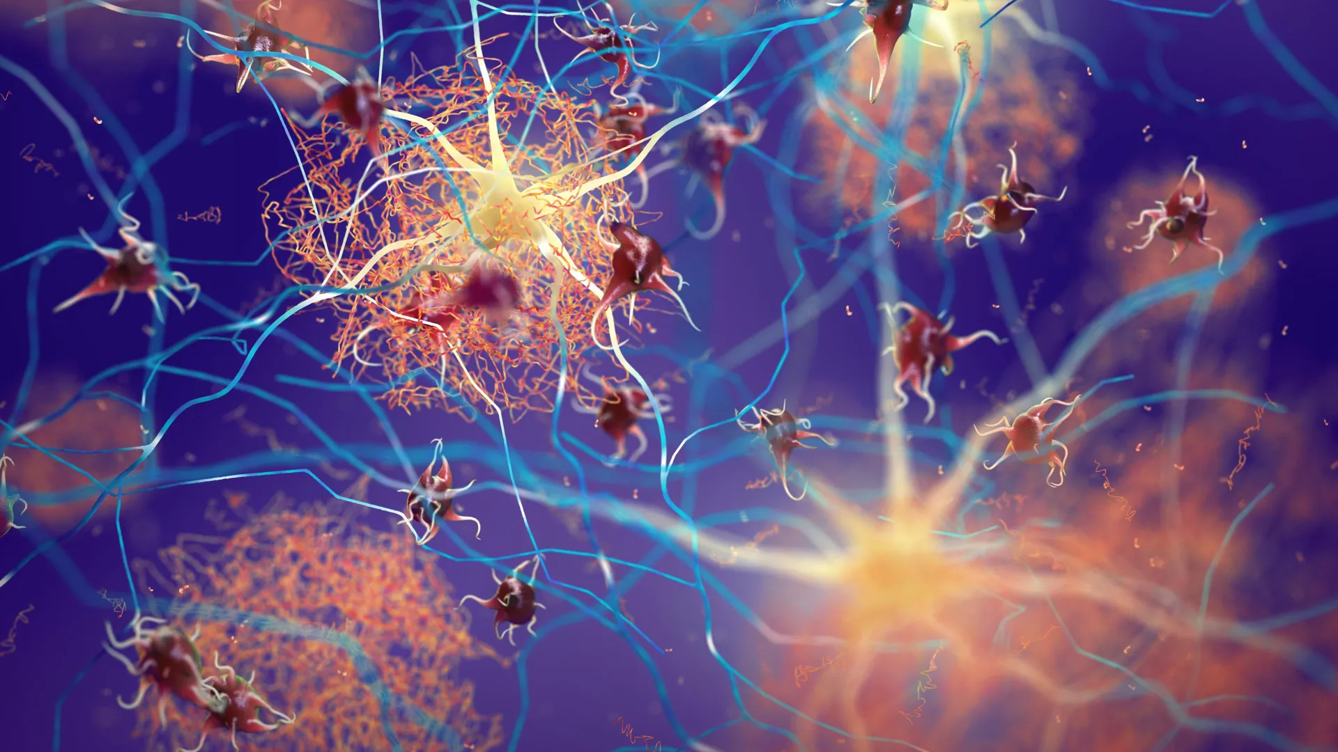
Alzheimer's disease is marked by the accumulation of harmful amyloid plaques that disrupt brain function. While many treatments focus on removing these plaques from the outside, a groundbreaking approach turns inward—harnessing the brain's own support cells to do the job. Scientists have discovered that by boosting a specific protein called Sox9, they can supercharge star-shaped cells known as astrocytes, which act as the brain’s maintenance crew. In mouse models that already exhibited memory deficits, this strategy significantly reduced plaque buildup and helped preserve cognitive performance over time. In this guide, we’ll walk through the step-by-step process researchers use to achieve this effect, from identifying the key protein to observing long-term benefits.
What You Need
- Animal model: Mice that have been genetically engineered to show Alzheimer's-like symptoms (e.g., APP/PS1 transgenic mice) – critical for testing plaque reduction.
- Sox9 upregulation tool: A viral vector (e.g., AAV) that delivers a Sox9 gene into the brain, or a small molecule that increases Sox9 expression.
- Astrocyte-specific promoter: To ensure Sox9 is turned on only in astrocytes. A promoter like GFAP or Aldh1l1 is typically used.
- Stereotaxic injection setup: Precision equipment to inject the viral vector into the hippocampus or cortex.
- Behavioral testing apparatus: Morris water maze, Y-maze, or novel object recognition tests to assess memory and learning.
- Brain tissue processing tools: Cryostat, antibodies for immunofluorescence (e.g., anti-amyloid beta, anti-Sox9, anti-GFAP), and confocal microscope.
- Data analysis software: ImageJ for plaque quantification, GraphPad Prism for statistical analysis.
Step-by-Step Instructions
Step 1: Confirm Baseline Cognitive Deficits in the Mouse Model
Before any intervention, verify that your transgenic mice show measurable memory impairments. Perform a simple behavioral test, such as the Y-maze, which checks spatial working memory. Record the number of spontaneous alternations; mice with Alzheimer’s typically make fewer correct choices. This baseline ensures you can later compare the effect of Sox9 upregulation. See tips for reliable testing.
Step 2: Design and Prepare the Sox9 Delivery System
Construct an AAV (adeno-associated virus) vector carrying the Sox9 coding sequence under an astrocyte-specific promoter. Use a control vector (e.g., GFP) for comparison. Purify and titer the virus. It’s essential to have high titers (≥10^12 vg/mL) for efficient transduction. Include a tag like FLAG or HA to track Sox9 expression later.
Step 3: Perform Stereotaxic Injection
Anesthetize the mouse and place it in a stereotaxic frame. Use coordinates targeting the hippocampus (e.g., AP: -2.0 mm, ML: ±1.5 mm, DV: -1.5 mm from skull). Inject 1–2 µL of the AAV-Sox9 into each hemisphere at a slow rate (0.2 µL/min). Leave the needle in place for 2 minutes after injection to prevent backflow. Suture the wound and allow recovery.
Step 4: Allow Time for Transgene Expression
Wait 3–4 weeks for Sox9 to be expressed and accumulate in astrocytes. During this period, monitor the mice for general health and weight. If using a chemical inducer, administer it daily according to protocol.
Step 5: Conduct Post-Intervention Behavioral Tests
Repeat the Y-maze test and add a more demanding task like the Morris water maze. Train mice over 5 days with hidden platform; measure escape latency and probe trial performance. Compare Sox9-treated mice with controls. In successful experiments, treated mice show shorter escape times and spend more time in the target quadrant – evidence of preserved spatial memory.
Step 6: Examine Brain Tissue for Plaque and Astrocyte Changes
Sacrifice the mice and perfuse with PBS followed by paraformaldehyde. Section brains (40 µm thick) using a cryostat. Perform double immunofluorescence staining: one antibody against amyloid-beta (e.g., 6E10) to label plaques, and another against GFAP to mark astrocytes. Also stain for Sox9 (or its tag) to confirm transduction. Use a confocal microscope to capture z-stacks.
Step 7: Quantify Plaque Burden and Astrocyte Activation
Calculate the percentage of area covered by amyloid plaques in the hippocampus and cortex using ImageJ. Also count the number of astrocytes and measure the intensity of GFAP or Sox9 signal. Statistical testing (t-test or ANOVA) will reveal significant differences. Expected result: Sox9-treated mice have fewer and smaller plaques, and more robust astrocyte processes surrounding the remaining plaques.
Step 8: Correlate Cognitive Performance with Molecular Changes
Perform correlation analyses between plaque load, astrocyte coverage, and behavioral scores. A strong negative correlation between Sox9 levels and probe trial performance (i.e., higher Sox9 → better memory) reinforces the therapeutic potential. This step validates that boosting Sox9 is not just clearing plaques but also functionally preserving cognition.
Tips for Success
- Choose the right mouse model: Not all Alzheimer's models show robust plaque deposition. The APP/PS1 line is widely used and develops visible plaques by 6 months of age. For more severe pathology, consider 5xFAD mice.
- Optimize viral injection: Injecting too quickly or at high volume can cause tissue damage. Always test with a dye first. Use bilateral injections if possible to ensure coverage.
- Control for off-target effects: Sox9 is also expressed in neural stem cells. Use astrocyte-specific promoters (like GFAP) to limit expression. Include a control vector that overexpresses only GFP.
- Timing is crucial: In the original study, treatment started when mice already showed memory deficits (6 months old). This mimics a late intervention. If you start too early, you might miss cognitive decline. If too late, plaques may be too dense.
- Astrocyte activation can be neuroprotective: However, overactivation may cause gliosis. Monitor for any reactive morphology (hypertrophy, increased GFAP) that might indicate a harmful response.
- Combine with other markers: Besides Sox9, check for changes in Bace1 (a beta-secretase) or APP processing. Astrocytes can clear amyloid via release of proteases like neprilysin.
- Long-term follow-up: Assess mice at 1, 3, and 6 months post-treatment. The original study showed benefits over time. A single time point may miss dynamic changes.
- Reproducibility: Run at least two independent cohorts. Use littermates as controls to minimize genetic variability.
- Human translation caution: These steps are for preclinical research only. Boosting Sox9 in human astrocytes through gene therapy carries unknown risks. Always follow ethical guidelines for animal research.